Thursday, July 06, 2006
Taking A Close Look At HepB
June 26, 2006
Filed under: in the news...
Using some of the latest investigational techniques, scientists at the Scripps Research Institute have discovered pretty amazing functional morphology of the Hepatisis B virus:
Hepatitis B virions, also known as Dane particles, are approximately 40 nanometers in size, and the capsid is surrounded by a membrane envelope. While the structure of the hepatitis B capsid has been studied intensively in vitro, until this study little was known about the structure and assembly of native capsids present in infected cells in vivo, and even less was known about the structure of mature virions.
"We used cryomicroscopy and image analysis to examine the native structure of HBV [hepatitis B virus ] capsids from transgenic mice and virions isolated from patient blood samples," Yeager said. [Mark Yeager, M.D., Ph.D. is a professor at Scripps -ed.] "By rapidly freezing the samples we were able to use cryo-electron microscopy to image the particles while they were maintained at the temperature of liquid nitrogen--around--300° F--which preserves them in a state close to what exists in vivo. Image processing allowed us to derive 3-D maps that revealed for the first time how the outer lipid envelope interacts with the capsid shell. "
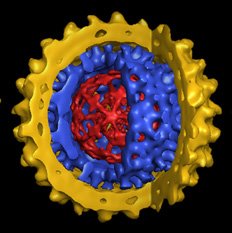
The 3-D maps showed that in terms of molecular size, hepatitis B virus is enormous--nearly 10 times larger than a hemoglobin molecule. Like the human genome, the genome of the hepatitis B virus is formed by double-stranded DNA and enclosed by the capsid, which has icosahedral symmetry, resembling the geometric structure of a geodesic dome. The capsid itself is contained within an outer envelope formed by a lipid bilayer, similar to the membranes that enclose all human cells. The membrane of hepatitis B virus is studded with glycoprotein spikes, which bind to receptors on liver cells that mediate infection.
In hepatitis B-infected liver cells, transcription of the viral DNA produces a type of RNA that is packaged into capsids. Within the capsid, reverse transcription produces a single-strand DNA copy that serves as the template for second strand DNA synthesis. The resulting particles bud through membranes of the endoplasmic reticulum--the part of the cell involved with protein folding, assembly, and transport--to acquire the outer membrane envelope of the virus, a step that confers infectivity.
"In the transgenic mice, we found two types of capsids with different densities," Yeager said. "While both types of capsids were assembled as similar icosahedral structures, we found that the lower density capsids did not contain any viral DNA or viral RNA, while the higher density capsids contained viral DNA intermediates. It seems likely that these lower density capsids were released from the cell nucleus. These results may offer new clues how the virus replicates in vivo."
# posted by Joyfulstill @ 9:24 PM 
![]()